BL1.2W: X-ray Tomographic Microscopy
The XTM beamline, located at BL1.2W, serves as a specialized facility at the Siam Photon Source (SPS) for X-ray Tomographic Microscopy (XTM) experiments. Utilizing the high-intensity X-ray beam from a 2.2-Tesla multipole wiggler, it provides researchers with advanced microtomography capabilities. This method allows for the reconstruction of cross-sectional details and the creation of 3D visualizations of diverse samples, with resolutions reaching as low as 1 micron (corresponding to a pixel size of 0.72 microns).
X-ray tomographic imaging, facilitated by the XTM beamline, is a powerful tool for researchers seeking a comprehensive 3D characterization of their samples. This non-destructive technique delves deep within the sample, revealing intricate details of its internal morphology, including the presence of engineering defects, porosity variations, and the intricate distribution of pores.
The XTM beamline welcomes researchers from both academia and industry to leverage its capabilities. Thai and international applicants can submit online proposals for beamtime access through the dedicated portal: https://user.slri.or.th/beamapp/index_home [Beamtime Application at SLRI].

Synchrotron radiation
X-ray tomographic microscopy (SRXTM): Unveiling Internal Structures
Synchrotron Radiation X-ray Tomographic Microscopy (SRXTM) is a powerful non-destructive imaging technique that utilizes the highly intense and tunable X-ray beam generated by a synchrotron light source. This X-ray beam is directed towards the sample of interest. The differential absorption of X-rays by the various components within the sample creates a characteristic transmission pattern.
To capture this pattern, a scintillator converts the transmitted X-rays into visible light. This light is then magnified by an objective lens system coupled to a high-resolution camera, resulting in a detailed X-ray image. For a complete 3D reconstruction, SRXTM acquires multiple X-ray images by rotating the sample through a specified angular range, typically at least 180 degrees. If the sample size exceeds the field of view of the detector, multiple scans may be necessary.
Following image acquisition, a series of calibration steps are performed. Flat-field correction, using both bright and dark current images, removes artifacts caused by camera sensitivity variations and optical path distortions. Preprocessing often includes noise reduction and beam intensity normalization to further enhance image quality. The preprocessed images serve as input for computational reconstruction. Employing the filtered back-projection algorithm, these images are transformed into sinograms, which are then used to generate the final 3D tomographic images. This collection of reconstructed slices, also referred to as tomograms, provides a comprehensive visualization of the sample's internal structure with high resolution in all three dimensions.


Applications of SRXTM
Synchrotron-based X-ray Tomographic Microscopy (SRXTM) is a powerful imaging technique used for characterization and assessment in various research fields. Here are some key points about SRXTM
Internal Details and 3D Visualization:
-
SRXTM allows researchers to reveal internal details of samples in three dimensions.
-
Computational reconstruction generates 3D images, enabling volume presentation of segmented features.
-
Unlike traditional thin-sectioning methods, SRXTM does not require the sample to be cut into thin slices.
Principles of SRXTM:
-
SRXTM relies on X-ray images resulting from differential absorption (absorption-contrast) and/or refractive index (phase-contrast) of the sample.
-
No staining or coating is needed, making it suitable for various applications.
Applications:
-
Biology, Entomology, and Paleontology:
-
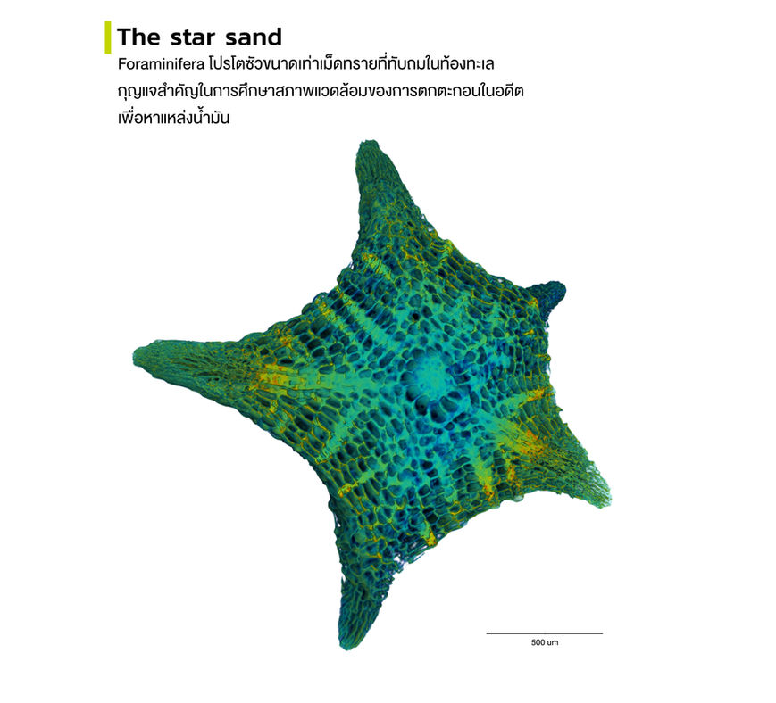
Useful for studying taxonomy, providing detailed morphology information about fruit seeds, small insects, and fossils.
-
Reconstructed images offer 3D visualization, allowing segmentation of internal features.
-
-
Life Science Research:
-
Widely used to examine porosity in animal models’ femurs, tibiae, and scaffolds.
-
-
Material Science Research:
-
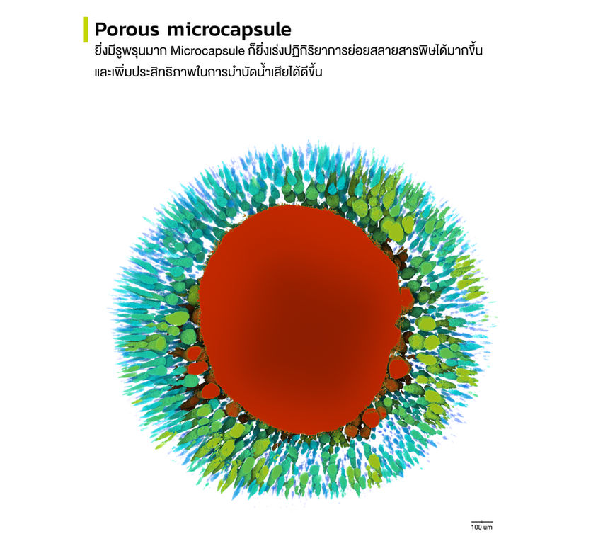
Investigates porosity in geopolymer and polymer microcapsules.
-
Presents pore distribution in 3D under different preparation conditions.
-
-
Food Processing:
-
Important for understanding pore distribution in processes like baking and roasting.
-
ABOUT US
TEAM STAFFS


CONTACT US
Mailing address:
Tomography and Imaging Section
Synchrotron Light Research Institute
Nakhon Ratchasima
30000, THAILAND
tomo.beamline
XTM Beamline


